
• Mon - Fri 8.00 - 17.30 & Sat 9.00-14.00

Blog

Categories Uncategorized

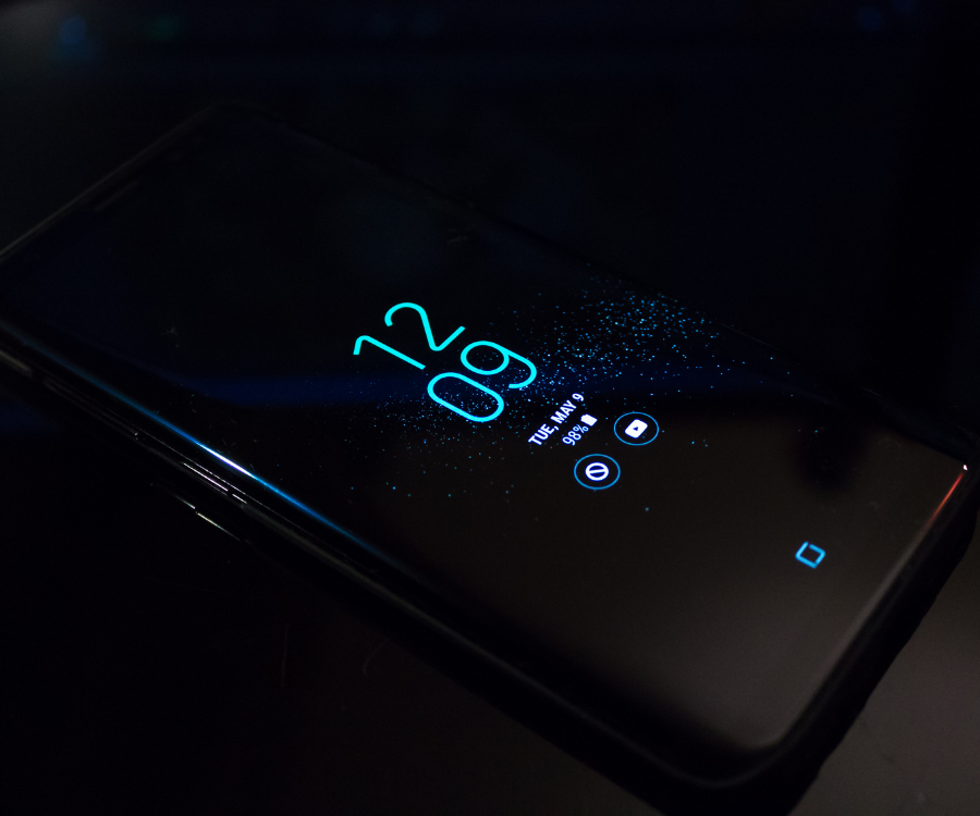
Can Dark Mode Help Your Eyes?

Can Dark Mode Really Save Your Eyes? What Science Actually Says

Every major operating system, browser, and app now offers a dark mode toggle. Many users flip the switch assuming it acts as an immediate shield against digital eye strain, late-night headaches, and insomnia.

The reality is more nuanced. While dark mode offers distinct advantages in specific environments, it is not an all-purpose cure for digital fatigue—and in some scenarios, it can actually make reading harder.

How Your Eyes Process Light vs. Dark Themes

Understanding the impact of dark mode requires looking at basic ocular optics:

  • Light mode (positive polarity): Dark text on a bright background. When looking at a light screen, your pupils constrict naturally in response to incoming illumination. Narrow pupils increase depth of field and sharpen optical focus, reducing the muscular effort needed to resolve letter shapes.
  • Dark mode (negative polarity): Light text on a dark background. In dimmer visual fields, pupils dilate to let in more light. Dilation decreases optical depth of field, which can introduce slight spherical aberrations and make edges look softer or hazier.

For high-speed reading comprehension and dense typographic work during daylight hours, the human visual system consistently performs better with positive polarity (dark text on light).

When Dark Mode Helps

Dark mode isn’t just an aesthetic trend; it provides measurable physiological and hardware benefits under specific conditions:

  • Low-Ambient Lighting: When browsing in a dark bedroom or a dimly lit workspace, an intense white display creates high contrast against the surrounding room. This stark difference forces retinal adaptation mechanisms to work continuously, causing discomfort. Dark mode harmonizes screen luminance with ambient darkness.
  • Melatonin Preservation: White displays emit substantial short-wavelength blue light, which triggers melanopsin-containing retinal ganglion cells that suppress melatonin production. Lower overall screen luminance at night helps maintain your body’s natural circadian cues.
  • Photophobia and Migraines: Individuals suffering from photophobia (light sensitivity), visual aura migraines, or post-concussion light sensitivity often experience immediate symptomatic relief by reducing overall luminous flux with dark interfaces.
  • Battery Conservation (OLED/AMOLED): On OLED screens, dark pixels are physically turned off rather than backlit. Running dark themes at low-to-moderate brightness significantly reduces display power consumption.

When Dark Mode Can Make Eye Strain Worse

Flipping on dark mode does not guarantee comfort, especially in these common scenarios:

1. The Halation Effect & Astigmatism

Roughly 30% to 50% of people have some degree of astigmatism—an irregular curvature of the cornea or lens that prevents light from focusing evenly on the retina.

When people with astigmatism view white text on a black background, dilated pupils cause light to bleed outward from letter strokes. This visual artifact, known as halation or the “halo effect,” makes text look blurry or shadowed, forcing the ciliary muscles inside the eye to work harder to maintain clarity.

2. Brightly Lit Workspaces

Using dark mode in an office flooded with daylight or overhead fluorescent lighting introduces reverse glare. Your eyes must continually adapt between the bright perimeter of your room and the dark rectangular canvas on your desk, triggering muscular fatigue.

Light Mode vs. Dark Mode: Direct Comparison

Visual FactorLight Mode (Dark Text / Light BG)Dark Mode (Light Text / Dark BG)
Best EnvironmentSunlit rooms, brightly lit officesDim rooms, nighttime, low-light flights
Reading SpeedHigher acuity and faster scanningSlightly slower text parsing speed
Visual AcuitySharper (pupils constrict)Softer; prone to halation/blurring
Astigmatism FriendlyYesOften worsens haloing around characters
Blue Light EmissionHigher overall lumen outputLower overall luminous output

What Actually Causes Digital Eye Strain?

If dark mode isn’t the silver bullet for computer vision syndrome (CVS), what is? Most symptoms blamed on “bright screens” actually stem from behavioral mechanics:

  • Reduced Blink Rates: Normal blinking occurs roughly 15 to 20 times per minute. Staring at digital tasks cuts that rate by half or more, leading to tear film evaporation, dry spots on the cornea, and stinging or burning sensations.
  • Fixed Focal Distance: Keeping eyes locked on a plane 50–70 cm away locks the ciliary muscles in continuous contraction, producing tension headaches and brow ache.
  • Improper Contrast & Glare: Reflections from windows or harsh overhead lighting washing out the display force extra visual effort to parse contrast.

4 Evidence-Based Habits for Healthier Eyes

Rather than relying solely on display palettes, adopt these proven ergonomic practices:

  1. Follow the 20-20-20 Rule: Every 20 minutes, shift your gaze to an object at least 20 feet away for 20 continuous seconds. This relaxes the ciliary muscle spasms responsible for focal fatigue.
  2. Match Display Luminance to Your Room: Hold a piece of white paper next to your screen. If the display looks like a lightbulb compared to the paper, it is too bright. If it looks gray or muddy, it is too dim.
  3. Use Gray Rather Than Pure Black: For reading light-on-dark, choose dark gray interfaces (such as #121212) rather than pure pitch black (#000000). Lowering contrast between the characters and the background reduces the intensity of the halation effect.
  4. Keep Artificial Tears on Hand: Preservative-free lubricating eye drops replenish the tear film disrupted by decreased blink frequency during long focus blocks.

The Verdict

Dark mode is a practical tool for nighttime use and low-light spaces, but it is not a cure for digital eye strain. For daytime reading, document editing, and users with astigmatism, standard light themes offer superior legibility and focus. Optimize your theme choice around your ambient room lighting rather than sticking to one permanent setting.

Categories Uncategorized

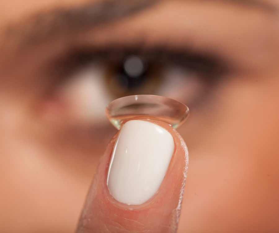
Contact Lenses: 10 Mistakes You Should Stop Making

Contact lenses offer freedom from frames, unobstructed peripheral vision, and everyday convenience. However, because they sit directly against delicate ocular tissue, improper use turns a medical device into a breeding ground for complications like corneal ulcers, chronic dry eye, and severe microbial infections.

1. Sleeping in Lenses Not Approved for Overnight Wear

Your cornea relies on atmospheric oxygen to stay healthy. Wearing contact lenses overnight starves the corneal surface of vital oxygen, causing corneal neovascularization (abnormal blood vessel growth) and increasing the risk of microbial keratitis up to eightfold. Unless your optometrist has prescribed extended-wear lenses, take them out before sleep.

2. Letting Water Touch Your Contacts

Showering, swimming, or rinsing lenses in tap water exposes your eyes to microscopic pathogens like Acanthamoeba—a waterborne amoeba that causes excruciating, vision-threatening infections.

  • Remove lenses before entering pools, hot tubs, or lakes.
  • Never store lenses in bottled, distilled, or tap water.
  • Keep eyes tightly shut if washing your face while lenses are in.

3. “Topping Off” Disinfecting Solution

Pouring fresh solution into old solution inside your lens case dilutes the active antimicrobial ingredients. This leaves residual bacteria, proteins, and lipids to incubate on your lenses overnight. Empty the case completely every morning, let it air dry upside down, and refill it with 100% fresh multipurpose or hydrogen-peroxide solution each night.

4. Keeping the Same Lens Case for Months

Contact lens cases develop invisible bacterial biofilms over time. Even if you scrub the plastic, bacteria embed themselves permanently. Replace your lens case every one to three months, or immediately whenever you open a new bottle of solution.

5. Stretching Replacement Schedules

Pushing a two-week disposable lens to four weeks or wearing monthly lenses for two months leads to protein buildup, reduced oxygen transmissibility, and micro-tears on the corneal epithelium. Adhere strictly to the manufacturer’s wear schedule, regardless of how comfortable the lenses still feel.

6. Skipping the “Rub and Rinse” Step

Even with “no-rub” multipurpose solutions, ophthalmological guidelines recommend gently rubbing your lenses with your fingertip for 5 to 10 seconds before rinsing. This physical action dislodges stubborn lipids, cosmetics, and environmental debris that chemical soaking alone cannot dissolve.

7. Touching Lenses with Wet or Dirty Hands

Transferring hand lotions, scented soaps, and residual moisture directly onto the lens causes stinging and transfers bacteria. Always wash your hands thoroughly with mild, fragrance-free soap, and dry them completely with a lint-free towel before handling lenses.

8. Wearing Contacts While Eyes Are Irritated

If your eyes feel gritty, red, or irritated, your eye is signaling distress. Inserting a contact lens traps irritants against the cornea and worsens inflammation. Remove the lenses immediately, switch to backup glasses, and consult an eye care professional if redness or pain persists beyond a few hours.

9. Applying Makeup Before Inserting Lenses

Powders, mascara flakes, and eyeliner pencils can easily smudge onto the lens surface if lenses are put in last. Follow this sequence:

  • Morning: Insert contacts before applying makeup.
  • Evening: Remove contacts before washing off makeup.

10. Skipping Routine Annual Eye Exams

Contact lens prescriptions require precise measurements of corneal curvature and tear film stability, not just visual acuity. Skipping annual checkups means missing early warning signs of hypoxia, asymptomatic infections, or improper lens fit before permanent corneal scarring occurs.

Common MistakeImmediate Eye RiskCorrect Practice
Tap water exposureAcanthamoeba keratitisMultipurpose or saline solution only
Topping off solutionIneffective disinfectionDiscard old solution daily
Over-wearing disposablesCorneal hypoxia, giant papillary conjunctivitisReplace on calendar schedule
Sleeping in lensesSevere bacterial keratitisRemove every night
Categories Uncategorized

Eye Test vs Vision Screening: What’s the Difference?

Eye Test vs. Vision Screening: What’s the Real Difference?

It is easy to assume that passing a school vision check or reading the line at the driver’s license bureau means your eyes have a clean bill of health. In reality, a quick eye screening and a comprehensive eye examination serve entirely different purposes.

Understanding the distinction between an eye test (comprehensive eye exam) and a vision screening is vital for protecting your long-term sight and overall ocular health.

What Is a Vision Screening?

A vision screening is a brief, preliminary check designed to identify potential, noticeable vision problems. It is not a diagnostic tool; rather, it acts as a filter to flag individuals who may need to see an eye care professional.

  • Who administers it: School nurses, pediatricians, primary care doctors, volunteers at health fairs, or licensing department officials.
  • What it tests: Basic distance visual acuity (often using a standard Snellen eye chart), gross eye alignment, and occasionally basic color perception.
  • The outcome: A simple result of “pass” or “refer.” A screening cannot diagnose why you are having trouble seeing or prescribe corrective lenses.
  • Limitations: Screenings frequently miss farsightedness (hyperopia), astigmatism, eye-teaming problems, focusing issues, and virtually all asymptomatic eye diseases.

What Is a Comprehensive Eye Test?

A comprehensive eye test is an in-depth medical evaluation of both your vision and the physiological health of your entire visual system.

  • Who administers it: A licensed optometrist or ophthalmologist.
  • What it tests:
    • Visual Acuity & Refraction: Pinpoints exact refractive errors (myopia, hyperopia, astigmatism, presbyopia) to generate an accurate optical prescription.
    • Binocular Vision & Alignment: Evaluates how well both eyes track, focus, coordinate, and perceive depth together.
    • Tonometry (Eye Pressure): Measures intraocular pressure to screen for glaucoma.
    • Retinal & Optic Nerve Assessment: Uses slit lamps, digital retinal photography, or dilation to inspect the retina, blood vessels, and optic nerve for conditions like macular degeneration, retinal detachment, or diabetic retinopathy.
    • Systemic Health Indicators: Detects early signs of broader health conditions, including diabetes, hypertension, high cholesterol, and autoimmune disorders.

Key Differences at a Glance

FeatureVision ScreeningComprehensive Eye Test
Primary GoalFlag potential distance vision issuesEvaluate total eye health and diagnose visual disorders
ProviderGeneral health worker, nurse, or trained volunteerLicensed optometrist or ophthalmologist
Scope of EvaluationBasic visual acuity (reading distant letters)Internal/external eye anatomy, eye pressure, binocular function
Prescription OutputNoneTailored prescription for glasses or contact lenses
Disease DetectionNoneDetects glaucoma, cataracts, macular degeneration, and systemic illness
Typical Duration2 to 5 minutes30 to 60 minutes

Why a “Pass” on a Screening Isn’t Always Enough

A common pitfall—especially in children—is assuming 20/20 distance vision equates to healthy eyes. A child can pass a school screening with 20/20 clarity while struggling with severe eye teaming or near-focus strain, leading to reading difficulties, headaches, and shortened attention spans in class.

Similarly, adult conditions such as glaucoma, diabetic eye changes, and early macular degeneration often develop silently without affecting distance clarity until permanent vision loss has occurred.

Categories Uncategorized

Why Blinking Less Could Be Damaging Your Eyes

Why Blinking Less Could Be Damaging Your Eyes (And How to Fix It)

Most people take blinking for granted. It happens automatically roughly 15 to 20 times per minute under normal conditions—amounting to over 14,000 involuntary wipes a day.

When staring at a computer monitor, smartphone, or television screen, that blink rate drops by up to 60%.

This phenomenon, often grouped under Digital Eye Strain or Computer Vision Syndrome, is more than just a minor inconvenience. Chronic under-blinking directly compromises the ocular surface and can trigger long-term vision and comfort issues.

What Actually Happens When You Blink

Blinking serves as the eye’s built-in maintenance system. Each full eyelid closure performs three critical functions:

  1. Spreads a Fresh Tear Film: Every blink coats the cornea with a microscopic three-layer shield consisting of mucin (to help tears adhere), aqueous water (to hydrate and deliver oxygen), and lipids (oils to prevent quick evaporation).
  2. Clears Debris: It washes away dust, allergens, and cellular waste into the tear ducts.
  3. Stimulates Oil Production: The mechanical squeeze of an eyelid activates the Meibomian glands along the eyelid margins, releasing the essential oils needed to seal the tear film.

When you stare intently at pixels, you do not just blink less often—you also perform more incomplete blinks, where the upper eyelid fails to touch the lower lid.

The Hidden Costs of Infrequent Blinking

ProblemPhysiological CauseCommon Symptoms
Evaporative Dry EyeLack of lipid renewal allows the aqueous tear layer to evaporate rapidly.Gritty sensation, burning, stinging, or redness.
Paradoxical Watery EyesDryness irritates corneal nerves, triggering an emergency flood of watery reflex tears that lack lubricating oils.Excessive tearing that fails to soothe dryness.
Meibomian Gland Dysfunction (MGD)Incomplete blinking leaves gland openings stagnant, causing oils to thicken and clog over time.Chronic irritation, crusty eyelid margins, inflammation.
Fluctuating Blurred VisionAn uneven, broken tear film distorts light before it even hits the cornea.Vision goes blurry while reading or working, clearing temporarily after a hard blink.

Practical Ways to Restore Healthy Blinking Habits

1. Master the 20-20-20 Rule

Every 20 minutes, look away from your screen at an object at least 20 feet away (around 6 meters) for 20 seconds. This relaxes the eye’s ciliary focusing muscle and naturally resets your baseline blink cadence.

2. Practice Deliberate Blink Breaks

If you work long hours on digital displays, perform intentional blink exercises twice a day:

  • Close your eyes normally for 2 seconds.
  • Open them.
  • Close your eyes again, gently squeezing the lids together for 2 seconds to express the meibomian oils.
  • Repeat 5 times per session.

3. Lower Your Screen Height

Position your monitor slightly below eye level (about 15 to 20 degrees). Looking slightly downward reduces the exposed surface area of the ocular globe, substantially cutting down the rate of tear evaporation.

4. Optimize Your Environment

  • Hydrate: Keep adequate water intake throughout the workday.
  • Humidify: Use a desktop humidifier if working in air-conditioned or heated rooms.
  • Redirect Airflow: Ensure desk fans, air conditioning vents, and car heaters are not blowing directly toward your face.
  • Use Preservative-Free Lubricating Drops: Artificial tears can provide barrier support during intensive screen sessions (avoid medicated “redness-reliever” drops for daily dryness).

If gritty sensations, burning, or intermittent blurriness persist despite adjusting your work habits, schedule an evaluation with an optometrist or ophthalmologist to check for underlying Meibomian Gland Dysfunction or chronic dry eye disease.